
Client Resources
The Cruciate Ligaments
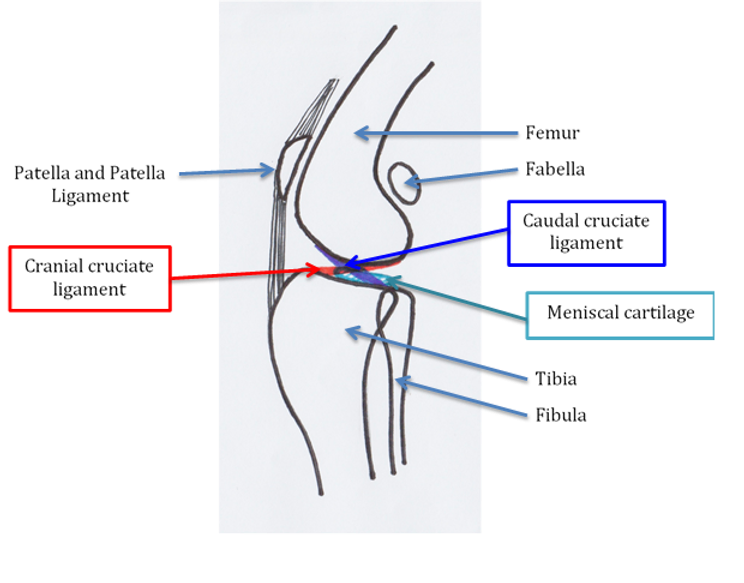
The cruciate ligaments are two structures within the stifle joint (the knee joint), the cranial cruciate ligament and the caudal cruciate ligament. They function to act against the forces of gravity and muscle movement, and create stability within the joint.
The caudal cruciate ligament originates from the front of the base of the femur, and inserts on the back of the top of the tibia. The cranial cruciate ligament originates from the back of the base of the femur, and inserts on the front of the top of the tibia. Together, these ligaments form a cross (hence the name cruciate).
Caudal cruciate ligament disease is very uncommon, but cranial cruciate ligament disease is considered one of the most common orthopaedic problems in dogs.
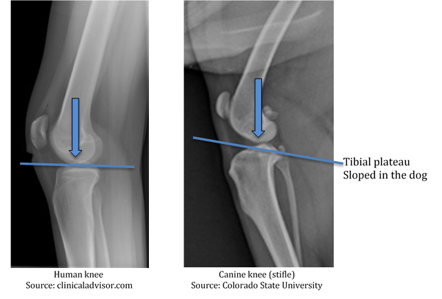
The knee joint in people functions quite differently to that of dogs. This is due to the fact that we stand upright (rather than on four legs), and the shape of our tibia. In people, the weight-bearing surface of the tibia is generally flat, creating a relatively stable platform when standing and walking, with the greatest forces on the joint exerted during running and sudden changes in direction. For this reason, cruciate ligament disease in people is most commonly due to trauma, often sports injuries.
In dogs, the tibial plateau is more sloped. This means that even when standing and walking, the knee joint is under directional force, causing the femur to move backwards down the slope of the tibial plateau, and the tibia to move forwards. The cranial cruciate ligament acts against these forces to keep the knee stable, and the femur and tibia firm in position relative to each other.
Degeneration of the cranial cruciate ligament can occur through several pathways. Excessive slope or angle of the tibial plateau can cause excess stress on the cranial cruciate ligament. The strength of the cruciate ligament fibres is often genetically controlled, but in some cases can be associated with autoimmune diseases. As these factors are a reflection of the dog’s conformation and genetic make-up, often these processes occur in both knees and get worse over time. CrCLD can occur in any dog of any age, but is more common in certain breeds such as Rottweilers, Staffordshire Bull Terriers, Mastiffs, Akitas, Labradors and Retrievers.
CrCLD can be a process of slow degeneration and partial tearing of the ligament, occurring over months or even years. Eventually this process can lead to complete tearing of the cranial cruciate ligament. This degenerative process is much more common than traumatic ligament rupture in an otherwise healthy ligament. However, traumatic ligament damage can occur in very active dogs.
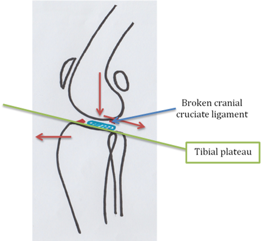
Once the cranial cruciate ligament is compromised, the forces within the knee joint go unchecked and the femur can slide down the back of the tibial plateau. This causes strain on the other supporting ligaments of the stifle, and excessive forces on the meniscal cartilages.
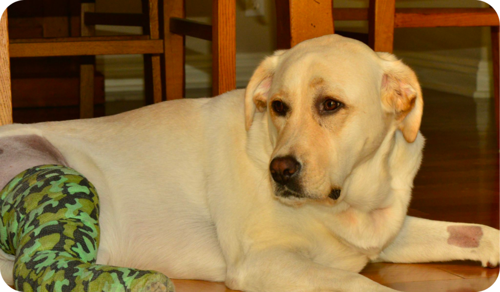
There can be a large variation in signs associated with CrCLD. The most common signs are limping, joint swelling, reluctance to play or run, stiffness, difficulty rising from sitting, altered sitting position, muscle loss over the affected limb(s), and clicking and crunching of the knee joint.
These can be slow changes noticed over time, or can occur suddenly.
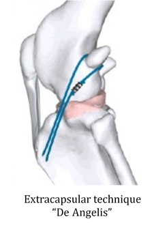
CrCLD is best managed with a surgical approach. There are several different surgical techniques. The most common techniques used are extracapsular repair (a.k.a. De Angelis), Tibial Tuberosity Advancement (TTA), Triple Tibial Osteotomy (TTO), and Tibial Plateau Levelling Osteotomy (TPLO).
Extracapsular repairs are commonly used in small dogs, usually less than 15kg. This technique is designed to mimic the action of the cranial cruciate ligament using an implant around the joint. The implant stimulates scar tissue to develop that provides additional support for the joint.
We are able to perform a De Angelis in our practice. Our estimates are generated for each individual, and they include an overnight stay in hospital, pre-anaesthetic blood testing, post-operative medications, and follow-up visits.
The TTA, TTO and TPLO techniques involve making cuts in parts of the tibia and modifying the angles of the tibial plateau or the patella ligament. These techniques aim to alter the directional forces in the knee joint.
The TTO and TPLO techniques are best performed by specialist orthopaedic surgeons in referral hospitals. They require high levels of training and specialised equipment to perform, but produce the best results. Referral for surgery minimises the risk of post-operative infection and complications. Referral is easily available, and estimates can be sourced upon request, but the average is $3500-$5000.
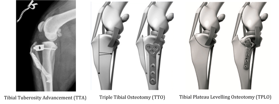
The TTA technique is available here in our practice, and our vets have undergone individual training to help give your pet the best outcome we can. You must remember, however, that while our vets have experience in this procedure and low rates of complications, they are not specialist orthopaedic surgeons. The TTA involves making an osteotomy (bone cut) along the front of the tibia, advancing it forwards and securing the adjustments with implants. This pulls the base of the patella ligament forwards and parallel to the direction of force through the joint. This force should be perpendicular to tibial plateau.
Our estimates are generated for each individual, and includes an overnight stay in hospital, pre-anaesthetic blood testing, post-operative medications, and follow-up visits and x-rays.
The meniscus is a crescent-shaped structure positioned on the tibial plateau within the knee joint. There are two in the stifle joint, one on the medial surface (towards the body midline), and one on the lateral surface (away from the body midline). They help to cushion the end of the femur and provide additional support and stability.
In cases of CrCLD, as the femur slides down the tibial plateau, the tibia can rotate inwards and cause uneven pressure on the medial meniscus, particularly the tail end. This excessive force can crush the medial meniscus and cause tears or displacement.
Meniscal injury is common with complete cruciate ligament rupture and is a source of significant pain.
The meniscal cartilages are inspected and assessed during the procedure for all knee surgeries. If the medial meniscus appears damaged, then the damaged portion can be removed to alleviate pain. In some cases, the whole of the medial meniscus is removed due to damage. While this does compromise stifle joint stability to a small degree, it is preferable to leave a damaged meniscus in the stifle that causes ongoing pain.
Damage to the meniscal cartilages can still occur even after surgery. These are called “late meniscal tears” and can occur in about 10% of cases. Some surgeons elect to perform a “meniscal release” to try to prevent late meniscal tears. This is done by cutting the attachment of the tail end of the medial meniscus, so it can freely move over the tibial surface and is less likely to be crushed by pressure. This however, also means the meniscus can’t perform its job as well as before. Your vet may give you the option to elect to perform a meniscal release as a preventative measure.
The most common complications of cruciate ligament surgery are late meniscal tear and infection. Infection can occur in up to 5% of cases, but we take several measures to minimise the chances of infection occurring. Many of the other complications that can occur are results of inappropriate post-operative care. Excessive or unrestricted activity, licking of the surgical site, and inappropriate physiotherapy can lead to joint swelling, persistent lameness, implant failure and, in some cases, fractures at the surgical site. Post-operative care is extremely important, and all efforts should be made to follow the recommendations made by your vet.
While all precautions are taken to ensure that your pet receives the best care possible, no procedure is without risk and sometimes the unexpected can occur. If you have questions regarding anaesthetic or surgical risk for your pet, please speak with our friendly staff.
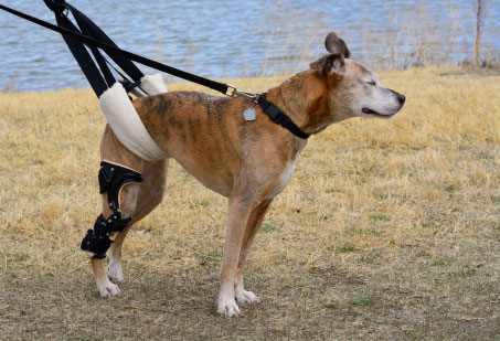
Each individual patient recovers at a different rate based on age, breed, weight, pre-existing disease, and age of the injury to the cruciate ligament. Generally speaking, the main post-operative recovery period is 8-12 weeks, with the first 4 weeks being the most intense. During the first 4 weeks, your pets must be strictly confined and supervised, so it is important that you have a suitable plan and set-up in place before your pet comes home after surgery. We recommend crate-resting your pet to minimise excessive activity, with short lead walks outside for toilet breaks. This does mean that a plan for keeping your pet happy during their rest period is needed, known as environmental enrichment. Your vet can give you a detailed recovery plan and advice about managing the initial rest period based on the needs of your pet.
If you have more questions about cruciate ligament disease, your pet’s diagnosis or treatment plan, please contact our friendly staff on 07 3297 0803 or come in and speak with us.