
Client Resources
Developmental Joint Disease
The term developmental joint disease refers to several types of joint disease that can affect the shoulders, elbows, stifles (knees), and hips in dogs. These diseases are characterised by abnormalities in cartilage structure, bone shape and length, or by fragments of bone interfering with joint movement. These diseases most commonly occur in medium to large breed dogs such as Great Danes, Mastiffs, Rottweilers, Labradors, Golden Retrievers, and German Shepherds, but many breeds can be affected. Dogs with developmental joint diseases will often show a persistent limp, stiff gait, difficulty getting up after resting or reluctance to play. These dogs will often show signs of joint pain before they are 2 years old, and may be affected on both sides of their body. The cause of developmental joint diseases is often poorly understood, but it is influenced by genetics and breed, and by nutrition during puppy hood.
OCD is a defect in the cartilage of a joint that may start as a crack in the cartilage. It may then progress to a flap, and further to a free-floating piece inside the joint. The cartilage defect causes pain and inflammation inside the joint. During an examination, your vet may detect pain on pressure over the affected joint, as well as when the joint is stretched and manipulated.
OCD lesions are sometimes visible on x-rays, but will sometimes require 3D imaging such as CT to be identified clearly.
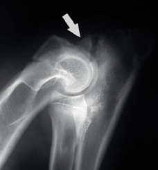
OCD lesions may occur in the shoulder joint, elbow joint or stifle (knee) joint. The x-ray image on the right shows an OCD lesion in the shoulder joint.
Surgery, or arthroscopy, is recommended to remove the cartilage defect, and often the long-term results are very good. The recovery after surgery for shoulder OCD lesions is excellent, often with full function after surgery. Significant improvement is often seen following surgery for elbow and stifle OCD lesions.
The Anconeal Process is part of the elbow joint that extends from the end of the ulna. It develops with a growth plate that normally closes or fuses as they approach 6 months of age. In some cases, the growth plate does not fuse normally and becomes ununited. An ununited Anconeal Process is slightly mobile, and causes instability of the elbow joint, pain and inflammation. During an examination, your vet may detect pain when the elbow is fully extended (straightened), and the elbow may not be able to flex (bend) completely.
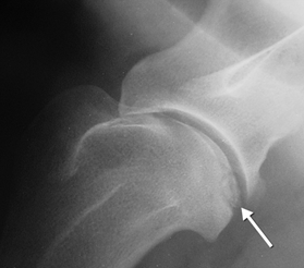
UAP may be diagnosed by x-rays of the elbow joint (as seen in the image on the left). The
recommended treatment for UAP is surgery, where the fragment is either removed, or is reattached to the ulna by screws or pins.
The Coronoid Processes are two small bony protrusions on the ulna within the elbow joint. Sometimes one of the Coronoid Processes will develop a crack and will separate from the ulna, creating a free-floating fragment. Similar to UAPs, the fragment causes instability of the elbow joint, pain and inflammation. During an examination, your vet may detect pain on pressure over the elbow joint, and the elbow may not be able to flex completely.
FCP is not visible on normal x-rays due to overlapping areas of bone causing superimposed lines. CT is needed to diagnose FCP, but we may assume a diagnosis of FCP based on the absence of other diseases on x-ray such as OCD and UAP.
Surgery is recommended to remove the fragment, and significant improvement is often seen following surgery.
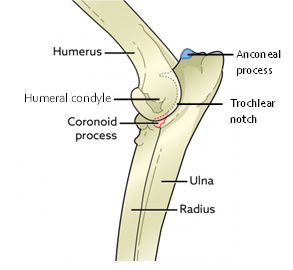
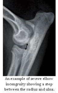
The elbow joint is formed by the humerus, the radius and ulna. All three must fit together exactly to create a normal joint. If one of these bones does not grow to fit the joint correctly, then an incongruity can occur. One form of incongruity is unequal growth between the radius and the ulna. Unequal growth may occur due to trauma or damage to one of the growth plates. Unequal growth of the radius and ulna may also affect the carpus joint (the “wrist”) and cause angular limb deformities (the limb appears twisted or bowed). Another form of incongruity is abnormal development of the shape of the trochlear notch in the ulna (in which the humeral condyles fit).
Joint incongruity can be identified on an x-ray, but may require CT to fully assess the shape of the bones and the joint. Surgery can be performed to help reduce the incongruity, but the recovery varies significantly between individuals due to the variation in the types of incongruity. Advice must be considered from a specialist veterinary orthopaedic surgeon.
Hip dysplasia is a common disease that may present at different times of an animal’s life depending on its severity. It is characterised by abnormal shape of the hip joint (formed by the pelvis and the femur), and results in instability of the joint known as “subluxation”. Subluxation causes the joint capsule and ligaments to stretch, causing pain and inflammation. Over time, subluxation causes abnormal wear on certain structures around the joint, which worsens the instability and inflammation. In severe cases, the instability can lead to dislocation of the hip joint. During an examination, your vet may detect pain on pressure over the hip joint, and pain on extension of the hip. The hip joint may not be able to extend completely.
Hip dysplasia may be identified on x-rays, but the signs seen on x-rays do not always directly relate to the painful signs seen in the animal.
Hip dysplasia may be managed in a range of different ways. Sometimes conservative treatment such as nutritional support and supplementation, and anti-inflammatory pain medications may be enough to manage the signs. Surgery may be recommended in some cases, with procedures such as Total Hip Replacements, or salvage procedures like Femoral Head & Neck Ostectomy.
Surgery for all of these diseases is best performed by a specialist orthopaedic surgeon. For some cases, we can arrange for a specialist surgeon to visit our clinic, but other cases will require referral to a specialist veterinary referral hospital such as Animal Referral Hospital, or Veterinary Specialist Services. For more information about surgical treatment and referral, please speak with your vet.